NOMIS Human Heart Atlas
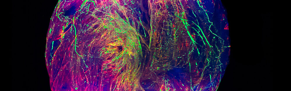
Biological tissues are organized in 3D from cells that differ in structure and molecular identity. Three-dimensional topographic cellular organization in whole organs is of paramount importance to their functions. Unique structural and molecular features of cardiac cells support essential heart function: beating about 100,000 times a day to pump 7,200 liters of blood to deliver a continuous supply of oxygen and other nutrients to the brain and the other vital organs. Until now, not much is known about the subtypes of cells in the human heart and how they are organized in 3D space. A high-definition spatial representation of cell types with their boundaries, neighbors, and interacting cells in the whole human heart is required to uncover the 3D network that is essential for sustaining life.
The NOMIS Human Heart Atlas: Charting the Cellular and Molecular Complexity of the Human Heart project aims to generate vascular, cellular and molecular maps of the entire human heart by merging novel technologies in imaging, spatial molecular profiling and artificial intelligence (AI). We aim to image and generate high-resolution maps of blood vessels and of various different cardiac cell types from the entire heart while revealing local proteomes from specific regions. In recent efforts, we rendered human organs transparent, enabling laser-scanning microscopy on intact human organs at the cellular level for the first time. Using deep-learning algorithms, we could analyze hundreds of millions of cells and entire vascular networks in large human organs, establishing the key technologies to aid the generation of high-resolution molecular and cellular maps of intact entire human organs.
The researchers anticipate that the maps they generate in this project can help to understand the cellular and molecular details of how human heart cells interact and function in physiological conditions, as well as during aging and in cardiovascular diseases.
The project is being led by Ali Ertürk at Helmholtz Zentrum München in Munich, Germany.
NOMIS researchers
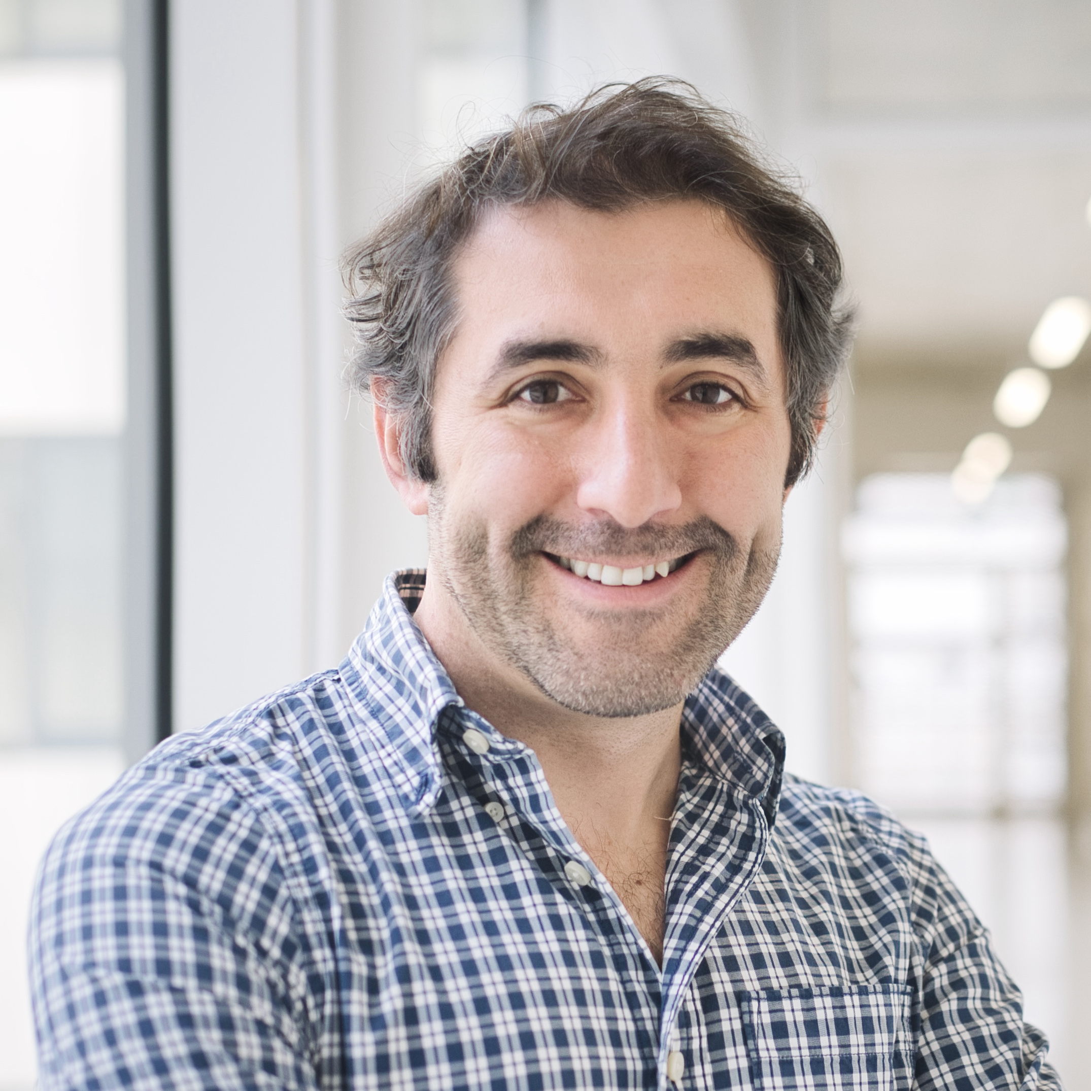
About Ali Ertürk Ali Ertürk is director of the Institute of Tissue Engineering and Regenerative Medicine (iTERM) at Helmholtz Zentrum München (Germany). He is leading the NOMIS Human Heart Atlas project. Ertürk studied molecular biology and genetics at Bilkent University in Ankara, Turkey, and obtained his PhD at the Max Planck Institute of Neurobiology in […]
Director of the Institute of Tissue Engineering and Regenerative Medicine
Helmholtz Zentrum München
Project Publications
Nanocarrier imaging at single-cell resolution across entire mouse bodies with deep learning
Efficient and accurate nanocarrier development for targeted drug delivery is hindered by a lack of methods to analyze its cell-level biodistribution across whole organisms. Here we present Single Cell Precision Nanocarrier Identification (SCP-Nano), an integrated experimental and deep learning pipeline to comprehensively quantify the targeting of nanocarriers throughout the whole mouse body at single-cell resolution. SCP-Nano reveals the tissue distribution patterns of lipid nanoparticles (LNPs) after different injection routes at doses as low as 0.0005 mg kg−1—far below the detection limits of conventional whole body imaging techniques. We demonstrate that intramuscularly injected LNPs carrying SARS-CoV-2 spike mRNA reach heart tissue, leading to proteome changes, suggesting immune activation and blood vessel damage. SCP-Nano generalizes to various types of nanocarriers, including liposomes, polyplexes, DNA origami and adeno-associated viruses (AAVs), revealing that an AAV2 variant transduces adipocytes throughout the body. SCP-Nano enables comprehensive three-dimensional mapping of nanocarrier distribution throughout mouse bodies with high sensitivity and should accelerate the development of precise and safe nanocarrier-based therapeutics.
Research Fields
Applied Sciences, Biomedical Engineering, Enabling & Strategic Technologies, Engineering, Nanoscience & Nanotechnology
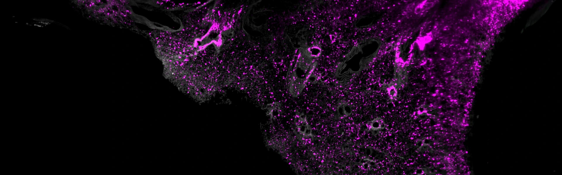
Persistence of spike protein at the skull-meninges-brain axis may contribute to the neurological sequelae of COVID-19
SARS-CoV-2 infection is associated with long-lasting neurological symptoms, although the underlying mechanisms remain unclear. Using optical clearing and imaging, we observed the accumulation of SARS-CoV-2 spike protein in the skull-meninges-brain axis of human COVID-19 patients, persisting long after viral clearance. Further, biomarkers of neurodegeneration were elevated in the cerebrospinal fluid from long COVID patients, and proteomic analysis of human skull, meninges, and brain samples revealed dysregulated inflammatory pathways and neurodegeneration-associated changes. Similar distribution patterns of the spike protein were observed in SARS-CoV-2-infected mice. Injection of spike protein alone was sufficient to induce neuroinflammation, proteome changes in the skull-meninges-brain axis, anxiety-like behavior, and exacerbated outcomes in mouse models of stroke and traumatic brain injury. Vaccination reduced but did not eliminate spike protein accumulation after infection in mice. Our findings suggest persistent spike protein at the brain borders may contribute to lasting neurological sequelae of COVID-19.
Research Fields
Biology, Biomedical Research, Clinical Medicine, Health Sciences, Immunology, Molecular Biology, Natural Sciences, Virology
Deep 3D histology powered by tissue clearing, omics and AI
To comprehensively understand tissue and organism physiology and pathophysiology, it is essential to create complete three-dimensional (3D) cellular maps. These maps require structural data, such as the 3D configuration and positioning of tissues and cells, and molecular data on the constitution of each cell, spanning from the DNA sequence to protein expression. While single-cell transcriptomics is illuminating the cellular and molecular diversity across species and tissues, the 3D spatial context of these molecular data is often overlooked. Here, I discuss emerging 3D tissue histology techniques that add the missing third spatial dimension to biomedical research. Through innovations in tissue-clearing chemistry, labeling and volumetric imaging that enhance 3D reconstructions and their synergy with molecular techniques, these technologies will provide detailed blueprints of entire organs or organisms at the cellular level. Machine learning, especially deep learning, will be essential for extracting meaningful insights from the vast data. Further development of integrated structural, molecular and computational methods will unlock the full potential of next-generation 3D histology.
Research Fields
Biochemistry & Molecular Biology, Chemistry
News
NOMIS researcher Ali Ertürk and fellow scientists at Helmholtz Munich, Ludwig-Maximilians-Universität (LMU) and Technical University Munich (TUM) have developed a technology that enables the precise detection of nanocarriers — tiny transport vehicles — throughout the entire mouse body at a single-cell level. The innovation could enable the targeted delivery of drugs, genes or proteins to cells for […]

In a Nature Methods perspective, NOMIS researcher Ali Ertürk addresses the new era of 3D-omics by tissue clearing and AI, called deep 3D histology. He writes that “biomedical research needs to evolve beyond the analysis of structural and molecular biology in selected tissue sections, expanding its focus to entire organs and organisms.” Ertürk is leading […]
NOMIS researcher Ali Ertürk and colleagues have developed a new chemical method, wildDISCO, that uses conventional antibodies and fluorescent markers to image a mouse’s entire body. This revolutionary technique provides detailed 3D maps that will enable a better understanding of biological systems and diseases. Their findings were published in Nature Biotechnology. More than a century […]