
Discovering the Causal Principles Underlying Brain-Wide Dynamics
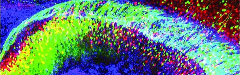

Light is a uniquely well-suited tool for probing living systems, which tolerate light as a natural part of the environment. Light can also be rapidly delivered and/or detected by experimentalists over a range of colors and powers, and in neuroscience can be used to both control (play-in) and detect (readout) neuronal activity patterns at the cellular level that give rise to behavior.
The Discovering the Causal Principles Underlying Brain-Wide Dynamics projects employed light to detect and understand brain states ― natural phenomena involving altered activity of brain cells that make the same experience mean completely different things to two different individuals, or even to the same individual at two different times. Such phenomena likely underlie not only personality differences but also neuropsychiatric disease states. However, it had been impossible to determine the nature of these states at the cellular level due to the immense complexity of brain structure and function.
To aid in this mission, the research team developed several methods, under conditions in which active neurons show fluorescence changes, to capture that light in real time across the brain to determine the natural activity patterns that mediate brain function and behavior. Accordingly, these new technologies were applied to allow the detailed identification of brain states: at cellular-resolution while maintaining a global, brain-wide perspective. Outcomes enabled a deeper understanding of ourselves, our similarities, our differences and our illnesses, at the most fundamental of levels.
NOMIS researchers

About Karl Deisseroth Karl Deisseroth is a 2017 NOMIS Awardee and a 2026 Nobel laureate. He is the D.H. Chen Professor of Bioengineering and of Psychiatry and Behavioral Sciences at Stanford University in the US and is an Investigator of the Howard Hughes Medical Institute. He led the project Discovering the Causal Principles Underlying Brain-Wide […]
D.H. Chen Professor of Bioengineering and of Psychiatry and Behavioral Sciences
Stanford Medicine
Project publications

Orbitofrontal cortex control of striatum leads economic decision-making
Animals must continually evaluate stimuli in their environment to decide which opportunities to pursue, and in many cases these decisions can be understood in fundamentally economic terms. Although several brain regions have been individually implicated in these processes, the brain-wide mechanisms relating these regions in decision-making are unclear. Using an economic decision-making task adapted for rats, we find that neural activity in both of two connected brain regions, the ventrolateral orbitofrontal cortex (OFC) and the dorsomedial striatum (DMS), was required for economic decision-making. Relevant neural activity in both brain regions was strikingly similar, dominated by the spatial features of the decision-making process. However, the neural encoding of choice direction in OFC preceded that of DMS, and this temporal relationship was strongly correlated with choice accuracy. Furthermore, activity specifically in the OFC projection to the DMS was required for appropriate economic decision-making. These results demonstrate that choice information in the OFC is relayed to the DMS to lead accurate economic decision-making. © 2023, The Author(s).
Research Fields
Biomedical Research, Developmental Biology, Health Sciences
Cardiogenic control of affective behavioural state
Emotional states influence bodily physiology, as exemplified in the top-down process by which anxiety causes faster beating of the heart1–3. However, whether an increased heart rate might itself induce anxiety or fear responses is unclear3–8. Physiological theories of emotion, proposed over a century ago, have considered that in general, there could be an important and even dominant flow of information from the body to the brain9. Here, to formally test this idea, we developed a noninvasive optogenetic pacemaker for precise, cell-type-specific control of cardiac rhythms of up to 900 beats per minute in freely moving mice, enabled by a wearable micro-LED harness and the systemic viral delivery of a potent pump-like channelrhodopsin. We found that optically evoked tachycardia potently enhanced anxiety-like behaviour, but crucially only in risky contexts, indicating that both central (brain) and peripheral (body) processes may be involved in the development of emotional states. To identify potential mechanisms, we used whole-brain activity screening and electrophysiology to find brain regions that were activated by imposed cardiac rhythms. We identified the posterior insular cortex as a potential mediator of bottom-up cardiac interoceptive processing, and found that optogenetic inhibition of this brain region attenuated the anxiety-like behaviour that was induced by optical cardiac pacing. Together, these findings reveal that cells of both the body and the brain must be considered together to understand the origins of emotional or affective states. More broadly, our results define a generalizable approach for noninvasive, temporally precise functional investigations of joint organism-wide interactions among targeted cells during behaviour. © 2023, The Author(s).
Research Fields
Clinical Medicine, Health Sciences, Neurology & Neurosurgery

All-optical physiology resolves a synaptic basis for behavioral timescale plasticity
Learning has been associated with modifications of synaptic and circuit properties, but the precise changes storing information in mammals have remained largely unclear. We combined genetically targeted voltage imaging with targeted optogenetic activation and silencing of pre- and post-synaptic neurons to study the mechanisms underlying hippocampal behavioral timescale plasticity. In mice navigating a virtual-reality environment, targeted optogenetic activation of individual CA1 cells at specific places induced stable representations of these places in the targeted cells. Optical elicitation, recording, and modulation of synaptic transmission in behaving mice revealed that activity in presynaptic CA2/3 cells was required for the induction of plasticity in CA1 and, furthermore, that during induction of these place fields in single CA1 cells, synaptic input from CA2/3 onto these same cells was potentiated. These results reveal synaptic implementation of hippocampal behavioral timescale plasticity and define a methodology to resolve synaptic plasticity during learning and memory in behaving mammals. © 2022 The Author(s)
Research Fields
Biomedical Research, Developmental Biology, Health Sciences
Project news

Karl Deisseroth has been awarded the 2026 Nobel Prize in physiology or medicine for discoveries leading to optogenetics.
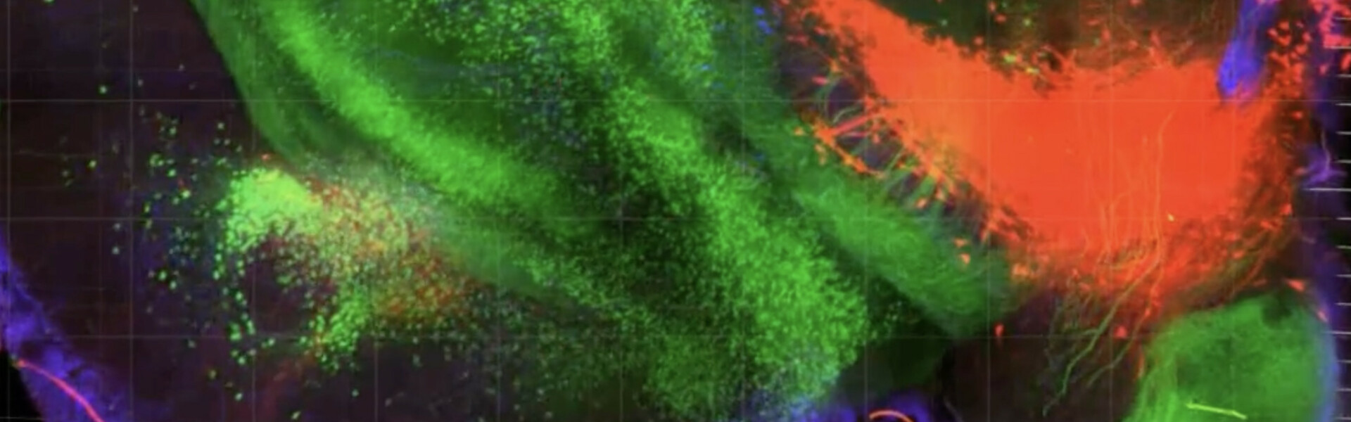

The NOMIS Foundation has released its third Insight film, which features 2017 NOMIS Awardee Karl Deisseroth and depicts the journey of his recently concluded NOMIS research project, Discovering the Causal Principles Underlying Brain-Wide Dynamics, which ran from 2017 to 2022. Created in collaboration with Vollformat, the film presents Deisseroth’s pioneering research that has generated new insights and tools […]


March 1, 2023
A racing heart drives anxiety behavior in mice
NOMIS Awardee Karl Deisseroth and colleagues have found that when they increased heart rates in mice, the animals showed more anxious behavior. Their research was published in Nature. Using pulses of light to control heart rate, Stanford Medicine researchers investigate a long-standing mystery about how physical states influence emotions. By Nina Bai Standing on the […]
