

Ali Ertürk
Director of the Institute of Tissue Engineering and Regenerative Medicine
Organization
Helmholtz Zentrum München

About Ali Ertürk
Ali Ertürk is director of the Institute of Tissue Engineering and Regenerative Medicine (iTERM) at Helmholtz Zentrum München (Germany). He is leading the NOMIS Human Heart Atlas project.
Ertürk studied molecular biology and genetics at Bilkent University in Ankara, Turkey, and obtained his PhD at the Max Planck Institute of Neurobiology in Munich, Germany. After five years of postdoctoral work at Genentech, South San Francisco, US, he returned to Munich as a principal investigator at the Ludwig Maximilian University of Munich in 2014. In 2019, he was appointed director of a new institute, the Institute of Tissue Engineering and Regenerative Medicine (iTERM), at Helmholtz Zentrum München.
His research aims to develop novel technologies based on tissue clearance and artificial intelligence (AI) to image and analyze intact rodent bodies, human organs, engineered tissues and organoids at cellular resolution. In contrast to the traditional histological methods relying on tissue sectioning and imaging of selected small regions, this new approach enables 3D imaging of the entire biological specimens at the cellular level at unprecedented speed and accuracy. Combined with molecular profiling, this hypothesis-free approach can substantially accelerate the understanding of complex and heterogenous biological systems.
‘s projects
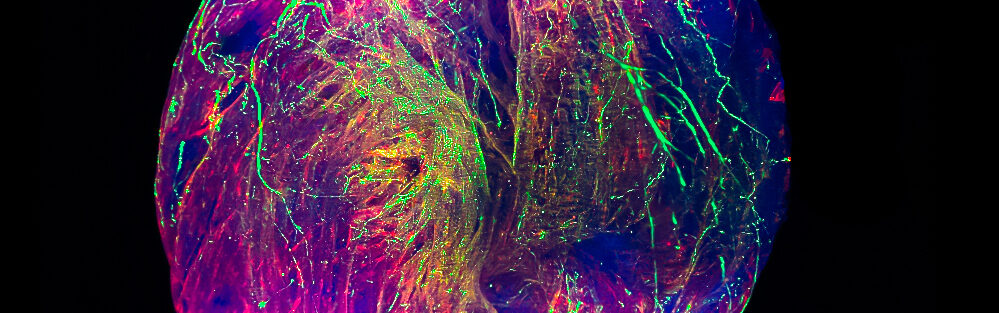
Biological tissues are organized in 3D from cells that differ in structure and molecular identity. Three-dimensional topographic cellular organization in whole organs is of paramount importance to their functions. Unique structural and molecular features of cardiac cells support essential heart function: beating about 100,000 times a day to pump 7,200 liters of blood to deliver […]
NOMIS researcher
Project period
2021 – 2027
‘s publications

A deep-learning framework reveals whole-body perturbations at cell level
Many diseases, including obesity, have systemic effects that perturb multiple organ systems throughout the body1,2. However, tools for comprehensive, high-resolution analysis of disease-associated changes at the whole-body scale have been lacking. Here we developed MouseMapper, a suite of foundation-model-based deep-learning algorithms enabling multi-system analysis of disease across the entire mouse body. MouseMapper enables whole-body quantitative analysis of nerves and immune cells, resolving fine axonal branches and immune-cell clusters while automatically segmenting 31 organs and tissues. We used MouseMapper to study diet-induced obesity, and identified structural alterations of the infraorbital branch of the trigeminal ganglia. This structural impairment in infraorbital nerves was associated with functional sensory deficits in whisker sensing. Furthermore, we identified proteomic changes in the trigeminal ganglion affecting axon remodelling and complement pathways both in mice and humans. MouseMapper also generated detailed three-dimensional inflammation maps by characterizing immune cell cluster compositions across tissues. The MouseMapper framework demonstrates robust generalizability across different imaging resolutions and datasets. Our study provides a powerful, scalable approach for identifying and quantifying systemic pathologies, bridging molecular insights from animal models to human conditions.
Research Fields
Clinical Medicine, Health Sciences, Neurology & Neurosurgery
A novel tamoxifen-inducible Mct8-CreERT2 mouse model for targeted studies of Mct8-expressing cells and thyroid hormone transport and function
Deficiency of the Monocarboxylate Transporter 8 (MCT8) severely impairs thyroid hormone (TH) transport into the brain, disrupting brain development as well as peripheral TH homeostasis. Studies assessing MCT8 expression patterns and tissue-specific pathologies induced by local TH-deficiency are often inconclusive due to unreliable antibody staining and the lack of functional tools to specifically target MCT8-expressing cells. For this purpose, we generated non-inducible Mct8-Cre and tamoxifen-inducible Mct8-CreERT2 mice. Mct8-Cre;Sun1-sfGFP mice demonstrated ubiquitous Sun1-sfGFP expression, due to early recombination driven by Mct8 gene expression at the stage of trophoblast implantation. Tamoxifen injection in 6-week-old Mct8-CreERT2 mice induced reporter expression specifically in Mct8-expressing cells in the brain and peripherally in liver, kidney, and thyroid, without leaky reporter expression in vehicle controls. Using vDISCO tissue clearing and 3D-imaging of GFP-nanobody-boosted mice, we further identified the sublingual salivary gland and the prostate as prominent Mct8-expressing organs. Nuclei from Mct8-expressing cells in the brain could selectively be enriched using fluorescence-activated nuclei sorting on Mct8-CreERT2;Sun1-sfGFP mice and characterized as choroid plexus cells and tanycytes. Our new inducible Mct8-CreERT2 line provides researchers with a tool to reliably mark, enrich, and characterize Mct8-expressing cells and to genetically modify genes specifically in these cells to study thyroid hormone transport and function.
Research Fields
Biomedical Research, Developmental Biology, Genetics & Heredity, Health Sciences
Nanocarrier imaging at single-cell resolution across entire mouse bodies with deep learning
Efficient and accurate nanocarrier development for targeted drug delivery is hindered by a lack of methods to analyze its cell-level biodistribution across whole organisms. Here we present Single Cell Precision Nanocarrier Identification (SCP-Nano), an integrated experimental and deep learning pipeline to comprehensively quantify the targeting of nanocarriers throughout the whole mouse body at single-cell resolution. SCP-Nano reveals the tissue distribution patterns of lipid nanoparticles (LNPs) after different injection routes at doses as low as 0.0005 mg kg−1—far below the detection limits of conventional whole body imaging techniques. We demonstrate that intramuscularly injected LNPs carrying SARS-CoV-2 spike mRNA reach heart tissue, leading to proteome changes, suggesting immune activation and blood vessel damage. SCP-Nano generalizes to various types of nanocarriers, including liposomes, polyplexes, DNA origami and adeno-associated viruses (AAVs), revealing that an AAV2 variant transduces adipocytes throughout the body. SCP-Nano enables comprehensive three-dimensional mapping of nanocarrier distribution throughout mouse bodies with high sensitivity and should accelerate the development of precise and safe nanocarrier-based therapeutics.
Research Fields
Applied Sciences, Biomedical Engineering, Enabling & Strategic Technologies, Engineering, Nanoscience & Nanotechnology
‘s news
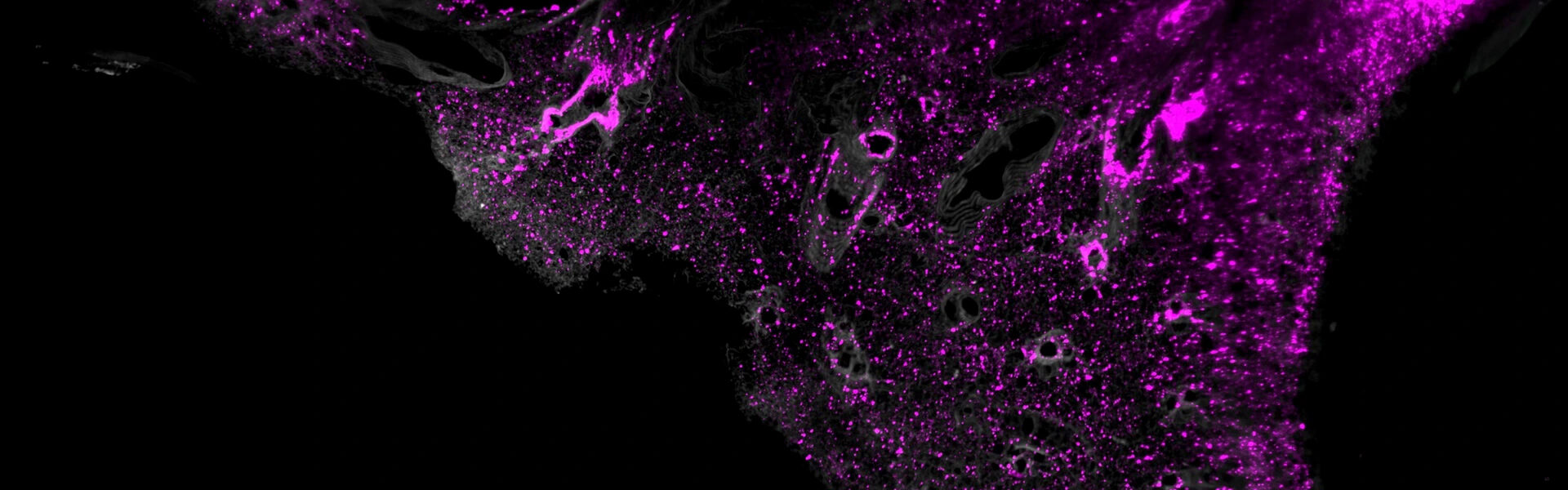

NOMIS researcher Ali Ertürk and fellow scientists at Helmholtz Munich, Ludwig-Maximilians-Universität (LMU) and Technical University Munich (TUM) have developed a technology that enables the precise detection of nanocarriers — tiny transport vehicles — throughout the entire mouse body at a single-cell level. The innovation could enable the targeted delivery of drugs, genes or proteins to cells for […]


In a Nature Methods perspective, NOMIS researcher Ali Ertürk addresses the new era of 3D-omics by tissue clearing and AI, called deep 3D histology. He writes that “biomedical research needs to evolve beyond the analysis of structural and molecular biology in selected tissue sections, expanding its focus to entire organs and organisms.” Ertürk is leading […]
NOMIS researcher Ali Ertürk and colleagues have developed a new chemical method, wildDISCO, that uses conventional antibodies and fluorescent markers to image a mouse’s entire body. This revolutionary technique provides detailed 3D maps that will enable a better understanding of biological systems and diseases. Their findings were published in Nature Biotechnology. More than a century […]
