Lung cancer hijacks immune cell metabolism to fuel its own growth
Published on
February 7, 2024
NOMIS Foundation Chair and director of the NOMIS Center for Immunobiology and Microbial Pathogenesis
Former NOMIS Fellow
Organization
NOMIS Center Director Susan Kaech and NOMIS Fellow Anna-Maria Globig, together with fellow scientists at the Salk Instistute, have discovered that lung adenocarcinoma cancer cells steer macrophage lipid metabolism to drive tumor progression, suggesting a new target for cancer treatment roadblocks.
Lung adenocarcinoma is the most common lung cancer and the cause of most cancer-related deaths in the United States. There are several ways lung adenocarcinoma can arise, one of which is a mutation in a protein called EGFR (epidermal growth factor receptor). Non-mutated EGFR helps cells grow in response to injury, but mutated EGFR promotes out-of-control growth that can turn into cancer. Modern immunotherapies don’t work against EGFR-driven lung adenocarcinoma, and while some drugs exist to treat the cancer, patients typically develop a resistance to them within just a few years. This gap in the treatment toolchest inspired Salk Institute researchers to probe for weak spots in the cancer’s growth pathway.
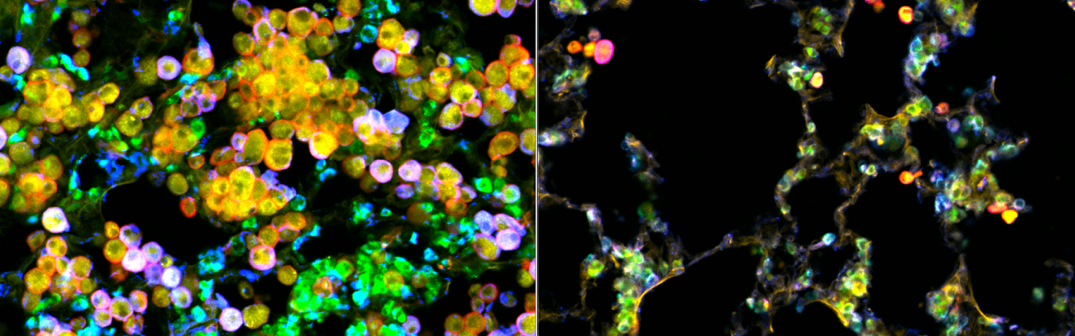
The team discovered that EGFR-driven lung adenocarcinoma hijacks a specialized population of lung-resident immune cells called macrophages, which are designed to dispose of diseased and damaged cells, as well as maintain a delicate balance of protective lipids (fats) around lung alveoli, which are essential for breathing. The lung cancer cells pull macrophages into the tumor microenvironment and alter their lipid metabolism to turn them into cancer fuel-suppliers. The newly energized tumor cells then spur further macrophage proliferation to supply more fuel—a novel self-perpetuating cancer mechanism.
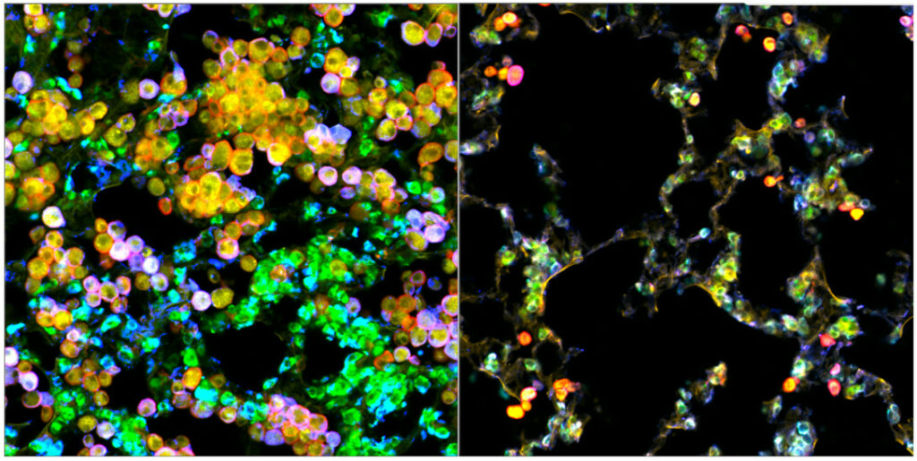
The findings, published in Cancer Discovery on January 25, 2024, provide new inspiration for lung adenocarcinoma interventions that disrupt this tumor cell-macrophage relationship. The researchers suggest that treatments using EGFR inhibitors may be more successful when paired with statins, a class of drugs commonly used to lower cholesterol levels.
“We have discovered a novel way that lung cancer cells manipulate their local environment and other cell types surrounding them to promote their own growth. In this case, the tumor cells exploit lung-resident macrophages—remodeling them to provide nutrients, like cholesterol, to the cancer cells and stimulate tumor growth,” says senior and co-corresponding author Susan Kaech, professor, director of the NOMIS Center for Immunobiology and Microbial Pathogenesis, and holder of the NOMIS Chair at Salk. “One exciting implication of this work is that lung cancer treatments may be improved by simply adding statins, an already widely used class of drugs, to the patient’s treatment plan.”
Continue reading this Salk Institute release
Read the Cancer Discovery publication: EGFR-Driven Lung Adenocarcinomas Co-opt Alveolar Macrophage Metabolism and Function to Support EGFR Signaling and Growth
NOMIS researchers
About Anna-Maria Globig Anna-Maria Globig is a NOMIS Center Postdoctoral Fellow at the Salk Institute for Biological Studies (La Jolla, USA). Globig was born and raised in Germany and obtained her medical degree from the Albert-Ludwigs-University in Freiburg im Breisgau. During her doctoral thesis, she investigated the immunological pathogenesis of inflammatory bowel disease. After receiving […]
Former NOMIS Fellow
Salk Institute for Biological Studies
About Susan Kaech Susan Kaech is the NOMIS Foundation Chair and director of the NOMIS Center for Immunobiology and Microbial Pathogenesis at the Salk Institute for Biological Studies (La Jolla, US). She co-led the Marmosets as a Model System of Aging and Neurodegeneration project and is currently co-leading the Neuroimmunology Initiative at the NOMIS Center. […]
NOMIS Foundation Chair and director of the NOMIS Center for Immunobiology and Microbial Pathogenesis
Salk Institute for Biological Studies